Breadcrumbs
- Home
- About Us
- Latest News
- MI at CAR 2025
MI at CAR 2025
The Canadian Association of Radiologists (CAR) 2025 Annual Scientific Meeting took place over the weekend in Montreal, Quebec from April 3-6, and several faculty and trainees represented the Department of Medical Imaging with their incredible abstracts, presentations and educational exhibits.
See what exhibits, abstracts and educational sessions MI presented:
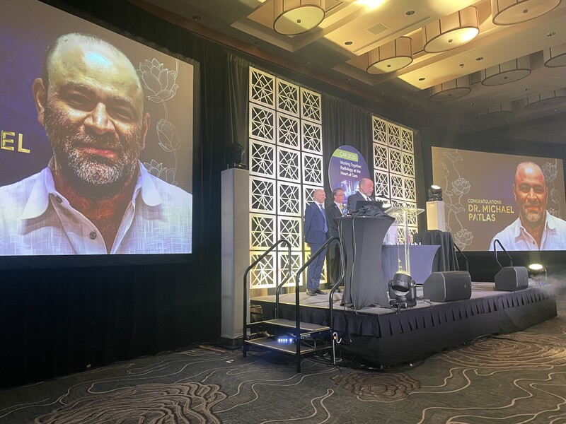
Dr. Michael Patlas, Chair, Medical Imaging
Refresher course title: Lessons Learned in the First Year as Chair
Dr. Patlas also delivered two presentations to the CAR Board & Canadian Heads of Academic Radiology on the Current Status of the CARJ, and chaired two meetings: Executive of Canadian Emergency, Trauma and Acute Care Society Executive; and the CARJ Editorial Board.
Award: Dr. Patlas, a long-time member of the CAR, also received the CAR Gold Medal Award to a standing ovation as for his numerous contributions to the Association and the radiology profession.
Dr. Kate Hanneman, VC, Research
Invited Talk title: Aortic Imaging Guidelines
Dr. Asutosh Sahu, Cardiothoracic Imaging Fellow, under supervision of Asst. Professor Dr. Binita Chacko
Abstract title: Radiologic features predicting local recurrence after Stereotactic Body Radiation Therapy in patients with primary lung cancer and lung metastasis.
Summary: The study results highlight the diagnostic challenge of distinguishing recurrence from treatment-related changes and suggest time-based cut-offs in follow-up imaging to improve accuracy.
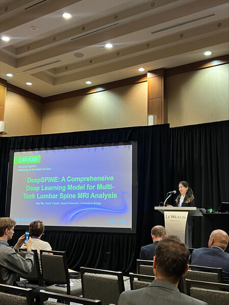
Dr. Kay Wu, PGY2 Diagnostic Radiology Resident
Study title: DeepSPINE: A Comprehensive Deep Learning Model for Multi-Task Lumbar Spine MRI Analysis
Summary: The DeepSPINE learning model was developed to automatically detect and grade degenerative spinal conditions from lumbar spine MRIs, and has shown promise in improving diagnostic accuracy, standardizing interpretations and streamlining workflows.
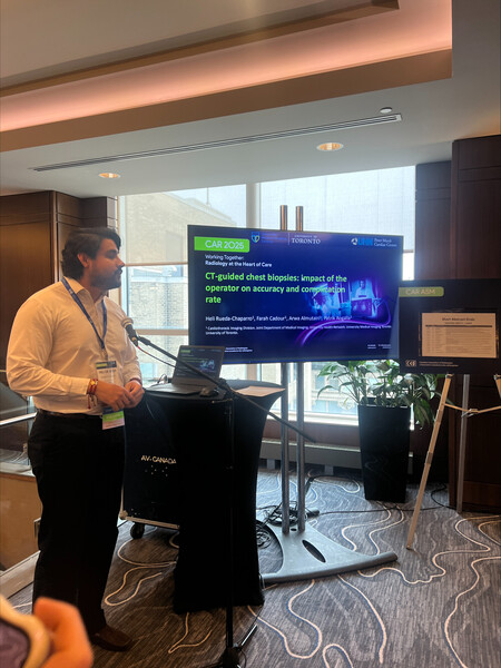
Dr. Heli Rueda Chaparro, Cardiothoracic Imaging Fellow, JDMI, under supervision of Dr. Patrik Rogalla, Cardiothoracic Imaging Division Head & Dr. Farah Cadour, Asst. Professor
Abstract title: CT-guided chest biopsies: impact of the operator on accuracy and complication rate
Summary: This study addresses the knowledge gaps in performing CT-guided chest biopsies.
Dr. Tiffani Ni, PGY1 Diagnostic Radiology Resident
Abstract 1: Imaging-Based Approach to Chronic Pelvic Pain: A Spotlight on Pelvic Venous Disease
Summary: This educational exhibit highlights Pelvic Venous Disease (PeVD) as an often underdiagnosed contributor to Chronic Pelvic Pain (CPP) and provides a structured approach to its imaging workup using ultrasound, CT, and MRI. By integrating key clinical and anatomical insights, this resource aims to enhance diagnostic accuracy and improve patient management.
Abstract 2: Pelvic Venous Disease: The Central Role of Radiologists in Diagnosis, Management, and Empowering Interdisciplinary Collaboration
Summary: This exhibit reviews the Symptoms-Varices-Pathophysiology (SVP) classification system, highlights the role of non-invasive imaging modalities like ultrasound, CT, and MRI, and discusses interventional radiology treatments such as endovascular embolization.
Abstract 3: Gaps in radiation safety education and what we can do about it: A creative radiation safety curriculum
Summary: Historic inadequacies in radiation safety education for medical trainees have led to knowledge gaps and suboptimal adherence to safety guidelines in interventional settings. To address this, the CAIR Radiation Safety Subcommittee has developed a multi-model curriculum to improve radiation safety and reduce radiation exposure in angiography suites, including an educational video on radiation safety in the Interventional Radiology suite for residents and fellows. Watch it here.
Dr. Arwa Almutair, Cardiothoracic Imaging Fellow, JDMI, with co-fellow Dr. Heli Rueda Chaparro, under the supervision of Dr. Patrik Rogalla, Professor & Asst. Professor Dr. Sonja Kandel
Abstract title: CT-fluoroscopy guided biopsy of lung lesions: Diagnostic sampling and pneumothorax rate for targets smaller vs. larger than 1 cm
Summary: This research assesses the risks and benefits of CT guided biopsy of small nodules less than 1 cm.
Dr. Jônatas Fávero Prietto dos Santos, Thoracic Imaging Fellow (graduated 2024), JDMI, under the supervisions of Dr. Patrik Rogalla, Cardiothoracic Imaging Division Head, Dr. Farah Cadour, Asst. Professor, and Dr. Felipe Sanchez, Asst. Professor
Abstract title: Improving post-lung biopsy care: CT chest at chest X-ray dose
Award-winning presentation: Second Best Radiologist-in-Training research project presentation
Summary: In this research conducted at Toronto General Hospital, titled "Improving post-lung biopsy care: CT chest at X-ray dose," we have demonstrated that ultra-low dose CT (ULDCT) chest, performed with advanced imaging techniques, offers superior diagnostic accuracy, improved image quality and interobserver agreement, and - most notably - radiation doses comparable to or even lower than those of a single standard chest X-ray. The advanced techniques used by our group included deep learning reconstruction (DLR) algorithms, silver beam filtration, and noise-mitigating weighted projections (thoracic tomogram), all combined to reduce image noise and improve image quality. Thoracic tomogram, an in-house post-processing technique derived from the ULDCT images that provides 2 cm thick-slices, helps to reduce reading time and further mitigate image noise. We are now entering a new era in which achieving chest CT at dose parity with a single chest X-ray is both accurate and feasible.
Dr. Sabreena Moosa, PGY2 Diagnostic Radiology Resident
Poster 1: Constipation on Radiographs: More than Just a Gut Feeling
Summary: This educational exhibit highlights an approach to reporting radiographs for the diagnosis of constipation and the differential diagnostic considerations and the terminology to provide the appropriate clinical direction when reporting radiographs for the diagnosis of constipation. Authors: Sabreena Moosa, Ania Kielar, Dilkash Kajal, Nasir Jaffer
Poster 2: Liver Transplant Patients with Benign Biliary Structures
Summary: Radiologist in training project
Authors: Sabreena Moosa, Eran Shlomovitz, Cathal O’Leary
Congratulations to all the Medical Imaging faculty and trainees who presented their work at the CAR 2025 Annual Scientific meeting and congratulations to all those who received awards! If you presented any work at the scientific meeting or received any awards for your work and you have not yet shared them with the Department and would like to, please email mi.communications@utoronto.ca with the following information and we will share your accomplishments in our MI Weekly newsletter and on social media:
- Headshot
- Your name, title, rank, division, site, residency/fellowship
- Names of faculty supervisor(s), if applicable
- Title of abstract/presentation/exhibit
- Brief 1-2 sentence summary of your work
- Name of award (if applicable)
- Twitter/Bluesky handle (if you’d like to be tagged*)
*Please note, if you’d like to be tagged in social posts on the Department’s LinkedIn page, you must be following the page here.
MI at the CAR 2025 - in photos
