Abdominal Imaging

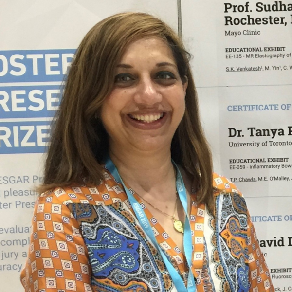
Dr. Tanya Chawla - Division Lead
Abdominal Imaging Fellowships Offered
The Division of Abdominal Imaging comprises faculty members from all of the university-affiliated teaching hospitals of the University of Toronto. These hospitals include:
- Sunnybrook Health Sciences Centre (SHSC)
- St. Michael's Hospital, Unity Health Toronto (SMH)
- Mount Sinai Hospital (MSH), Women’s College Hospital (WCH), and the University Health Network (UHN -- Toronto General Hospital, Toronto Western Hospital, and Princess Margaret Cancer Centre)
The division is organ-based, with most staff involved in multimodality imaging of the abdomen and pelvis. The modalities include the conventional techniques of plain film radiography, fluoroscopy examinations, barium examinations, hysterosalpingography, and nuclear medicine as well as the cross-sectional imaging modalities of ultrasound, computed tomography, and magnetic resonance imaging. Some teaching hospitals have their unique focus in abdominal imaging. For example, SHSC and SMH are both large trauma centres and are the only accredited Level I trauma centres in Canada. SHSC and UHN are both large cancer centres. UHN is the largest transplant centre in Canada. SMH has a strategic focus on radiology AI tool development. MSH is a major obstetrical ultrasound centre. The Abdominal Imaging Division leads research, teaching and clinical utilization on cutting-edge abdominal imaging techniques such as contrast-enhanced ultrasound, CT colonography, dual energy CT, prostate MRI, MR elastography, CT/MR enterography, and many more. The goal of the division is to achieve excellence in clinical work, teaching, and research.
Abdominal imagers are an integral part of patient care. Staff and fellows have close communication with the clinicians, are involved in numerous clinical rounds, and are present at all tumor boards.
Abdominal Education
Faculty members actively participate in teaching at the fellowship, resident, and undergraduate levels and also contribute to many CME programs at local, national, and international venues.
The University of Toronto offers several one-year clinical or two-year clinical research fellowships. Abdominal fellows are selected annually. These positions offer varying exposure to the various organ systems and modalities, depending on the hospital. A wide exposure to all areas is available. Fellows are encouraged to actively partake in teaching and research and to attend the many teaching rounds, lectures and Journal Club. Fellows are given protected research time and are expected to publish their research during the fellowship.
The University of Toronto has a large Residency Training Program in Diagnostic Imaging. One- to three-month rotations through all the Abdominal Imaging services are enhanced by daily teaching rounds at the affiliated hospitals, a lecture series in the PGY2-PGY3 years, and easy access to large hospital teaching files and the American College of Radiology (ACR) teaching file. Review lectures, mock oral examinations, and OSCE examinations are also provided by faculty annually with additional sessions provided for the final year residents prior to the Board examination. Each clinical rotation has specific goals and objectives with increasing responsibility given to the residents as they progress through the training program.
Faculty members are also active in undergraduate teaching. Faculty use imaging to teach anatomy to medical students in a structured lecture series, while also providing a special lecture series introducing the specialty of Diagnostic Imaging. There is also a large elective program for medical students in the Department of Medical Imaging.
The Division of Abdominal Imaging, as the largest Division of Medical Imaging, provides a significant component of presentations at the annual University of Toronto Organ Imaging Review Course. The Division is also significantly involved in an annual OBGYN ultrasound course. Small group hands-on workshops are run in CT colonography and prostate MRI a few times annually.
Abdominal Research
Our staff are involved in both phenotypic and quantitative research including but not limited to Obstetrics and Gynecology, Hepatobiliary, Prostate MRI, contrast ultrasound, trauma, and inflammatory bowel disease. This includes both clinical and lab research, often with basic science researchers. Through the Research Program, our faculty are able to apply for protected research time.
All residents must complete one research project during their training and are given a half-day per week of protected research time. Residents, fellows and staff research are presented at an annual research day and many of these projects are subsequently presented and published at national and international levels.