Breadcrumbs
- Home
- About Us
- Latest News
- Meeting MI: Dr. Jônatas Fávero Prietto dos Santos
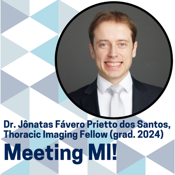
Meeting MI: Dr. Jônatas Fávero Prietto dos Santos

#MeetingMI offers the Department of Medical Imaging the opportunity to meet the members of MI and learn about their experience in their own words, starting with the next generation of radiologists: MI Residents and Fellows.
Name: Jônatas Fávero Prietto dos Santos
Residency Training Program name & PGY or Fellowship name: Thoracic Imaging Fellow (Graduated in 2024)
Hospital site: Joint Department of Medical Imaging (UHN, Sinai Health, Women’s College Hospital)
1. What inspired you to pursue radiology residency/fellowship?
I chose to pursue this fellowship to deepen my knowledge in thoracic and cardiac imaging. I had also always wanted to have a professional experience abroad. This year has been incredibly enriching—not only professionally, but also personally and culturally. Working at Toronto General Hospital and living in Toronto has been a turning point in my life. I’ve grown and learned so much!
2. What were the biggest highlights and challenges of your education in MI?
One of the biggest highlights of my fellowship was the opportunity to train at Toronto General Hospital, a high-complexity tertiary care center that offers fellows and residents a wide range of complex and fascinating thoracic and cardiac imaging cases. I spent nine months focused on thoracic imaging and three months dedicated to cardiac imaging, which gave me a comprehensive and well-balanced experience. Another major strength of the program is the cardiothoracic imaging division itself—made up of more than 20 staff radiologists. Being exposed to so many skilled professionals and learning from their different perspectives has shaped the way I approach and interpret studies. In addition, we gain extensive hands-on experience with transthoracic/percutaneous biopsies, which is a valuable aspect of the training.
As for challenges, adapting to a new healthcare system, hospital environment, and non-native language was certainly one of the biggest at first. However, the welcoming and supportive atmosphere created by the staff and co-fellows made this transition much smoother, allowing me to focus on the learning experience and get the most out of the fellowship.
3. You recently received an award at the CAR 2025 Annual Scientific Meeting for your research project; what does this award mean to you and your work?
This award was a very meaningful recognition of the collaborative work carried out by our team, led by Dr. Patrik Rogalla, with key contributions from Dr. Farah Cadour and Dr. Felipe Sanchez. It reinforces the value of conducting research with a clear goal of translating research findings into bedside care improvement.
On a personal level, being involved in this project strengthened my belief that it’s possible to achieve high-quality imaging even with ultra-low-dose radiation protocols. This has a direct impact on patient care by minimizing exposure to ionizing radiation, and it’s a principle I hope to apply and promote in my clinical practice moving forward.
4. What inspired this project and what significance are you hoping this research has in radiology?
There’s been growing interest in using ultra-low-dose radiation protocols in chest CT for different clinical scenarios, especially with the recent progress in advanced imaging techniques, like deep-learning reconstruction algorithms and the use of a silver beam filter during CT acquisition. Noise-mitigating weighted projections are also important, particularly a post-processing tool developed in-house called the Thoracic Tomogram, which also has contributed to improving image quality and work efficiency.
These combined innovations have made it possible to acquire high-quality ULDCT images at radiation doses similar to a standard chest X-ray. The research we’ve been doing in our department is helping to solidify this significant improvement in patient care.
Looking ahead, this could mean that, in certain situations, ULDCT might replace chest X-rays—offering better diagnostic accuracy at the same radiation dose. But we still need more research to understand how this could affect patient management and whether it makes sense economically in everyday clinical practice.
These techniques are also promising for patients who need repeated chest CTs, like oncology or immunocompromised patients. Recent studies, including from our department, have demonstrated that ULDCT can match the diagnostic accuracy of standard low-dose CT for detecting pneumonia, while reducing radiation dose even further.
5. What has your career been like since graduating from MI’s Thoracic Imaging Fellowship?
Since finishing the Fellowship, I’ve been living in Toronto, working on research projects with the team led by Dr. Rogalla, and wrapping up my PhD, which I’m doing through a university in Brazil. During this time, my wife has been completing her fellowship in liver transplant anesthesia at Toronto General Hospital, which finishes in June. After that, we’re moving back to our home country, and I’m very excited to apply the knowledge and experience I’ve gained here in my clinical and academic practice.
6. Tell us something about you that might surprise your colleagues!
Radiology wasn’t love at first sight for me. Since my second year of medical school, I focused on thoracic surgery, as my hometown, Porto Alegre, is a reference in this field in Latin America—much like Toronto is globally. For example, the first living-donor lung transplant outside the US was performed in Porto Alegre in 1999, and Toronto is known for its world-leading contributions to lung transplantation. I even started a general surgery residency, but soon realized it wasn’t the right fit for me. However, my exposure to chest X-rays and CT scans during thoracic surgery and pulmonology clinics, combined with my interest in radiologic anatomy, helped guide me towards radiology. Now, I’m extremely happy and fulfilled professionally in this field.